Swiss research
Swiss researchers find brain-structure differences linked to chronic pain
A new study from the Inselspital in Bern and the University of Fribourg finds structural differences in the cerebral cortex of people with primary chronic pain. The research, based on MRI scans of 30 chronic-pain patients and 30 healthy participants, adds evidence that persistent pain is associated with changes in the brain.
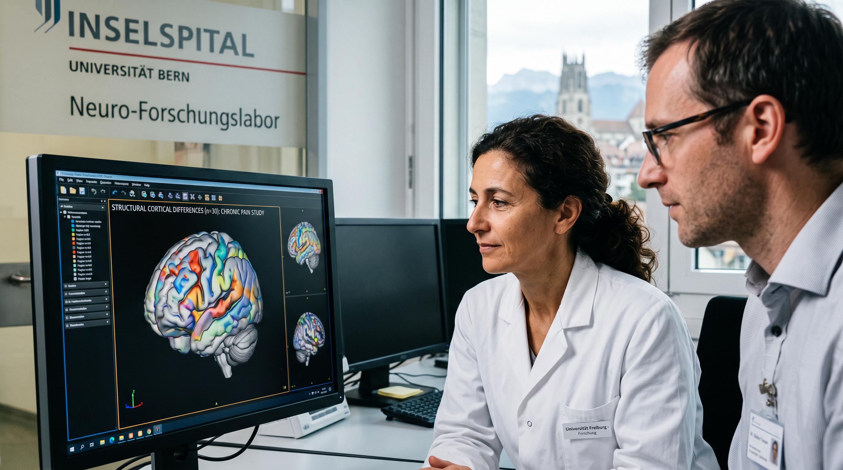
Swiss MRI Study Maps Pain’s Brain Changes
Sixty MRI scans are giving Swiss researchers a sharper view of chronic pain. A team from Inselspital Bern and the University of Fribourg has identified structural differences in the cerebral cortex of people living with primary chronic pain, according to a study funded by the Swiss National Science Foundation.
The researchers examined 30 people with primary chronic pain and 30 healthy participants using structural magnetic resonance imaging. Primary chronic pain describes conditions that cannot be fully attributed to a specific injury or disease. Fibromyalgia is a typical example.
The findings add to earlier evidence that persistent pain is associated with changes in the brain. They do not suggest that pain is imaginary or purely psychological. The scans point to physical differences in the outer layer of the brain, including areas involved in emotion, memory, sensation and pain assessment.
The study comes as clinicians continue to manage pain that outlasts the original trigger, often affecting work, sleep and daily life. Its results may help researchers refine how they understand the progression from short-term pain to a long-lasting condition. They do not yet provide a diagnostic scan or a new treatment for patients in Switzerland.
Researchers Find Distinct Cortical Folds
The left and right hemispheres showed different cortical patterns. In participants with primary chronic pain, a region at the front of the left hemisphere was more deeply folded than in the healthy comparison group. The cerebral cortex is the brain’s outer layer, shaped by folds and grooves that increase its working surface.
On the right side, researchers found shallower grooves in several areas, including regions toward the front of the brain. These areas help process sensory stimuli and pain. The left frontal region identified by the team also contributes to emotional and memory processes and has been associated with how people perceive and evaluate chronic pain.
The pattern matters because persistent pain involves more than the transmission of a signal from the body. Pain can affect attention, mood, memory and expectations, while stress and negative emotions can influence how the brain evaluates sensations. Salome Häuselmann of Inselspital Bern said the findings fit a hypothesis that emotional and cognitive processes become increasingly important as acute pain becomes chronic.
The researchers describe a possible reinforcing cycle involving pain signals, stress, emotions and cognitive assessments. The MRI results show an association, however, not proof that one factor directly causes another.
One Scan Cannot Settle the Cause
The study cannot yet say whether pain reshaped the brain or whether the brain increased pain vulnerability. Because the researchers examined participants at one point in time, the results provide a cross-section rather than a record of change.
That distinction is essential for patients and doctors. A structural difference on an MRI scan does not establish when it appeared, what caused it, or whether it predicts the severity of someone’s symptoms. The study also involved a relatively small group of 60 participants, so larger research will be needed to test whether the pattern applies across different forms of primary chronic pain.
Häuselmann said long-term studies would be necessary to answer the cause-and-effect question. Such research could follow people from an initial injury or episode of pain, track changes in symptoms and brain structure, and examine how treatment affects both over time.
The findings also reinforce the need to treat chronic pain as a condition involving the whole person. Medical care may need to address physical sensations alongside sleep, stress, mood, memory and the way patients interpret pain. That approach does not reduce pain to psychology. It reflects the interaction between brain systems that the Swiss study has begun to map.
Swiss Researchers Turn to the Next Scan
The researchers see room for the brain to adapt. Häuselmann said effective pain management might eventually be accompanied by cortical folds and grooves that more closely resemble those seen in people without chronic pain. Whether such structural changes occur, and whether they correspond to improved symptoms, remains unclear.
That uncertainty sets the next task for Swiss pain research. Future studies will need repeated MRI scans, larger participant groups and detailed clinical records. Researchers will also have to distinguish between different chronic pain conditions and account for treatments, sleep, stress and mental health, all of which can affect the experience of pain.
For people living with fibromyalgia and other primary chronic pain conditions, the findings offer a biological framework without promising a quick explanation or cure. The study supports the view that persistent pain has measurable effects across brain networks involved in sensation, emotion and memory. It also leaves open the possibility that those networks can change again.
Swiss hospitals and universities are likely to remain important in building that evidence. For now, the Bern and Fribourg team has produced a carefully limited result: chronic pain is associated with distinct cortical structure, while the direction and clinical meaning of that association still require years of follow-up research.