Switzerland
Zurich hospital uses hologram to guide brain surgery
Zurich University Hospital has performed brain surgery using mixed-reality glasses to project a three-dimensional hologram of the patient’s brain above the operating table. The technology is intended to help surgeons place a drainage catheter more precisely.
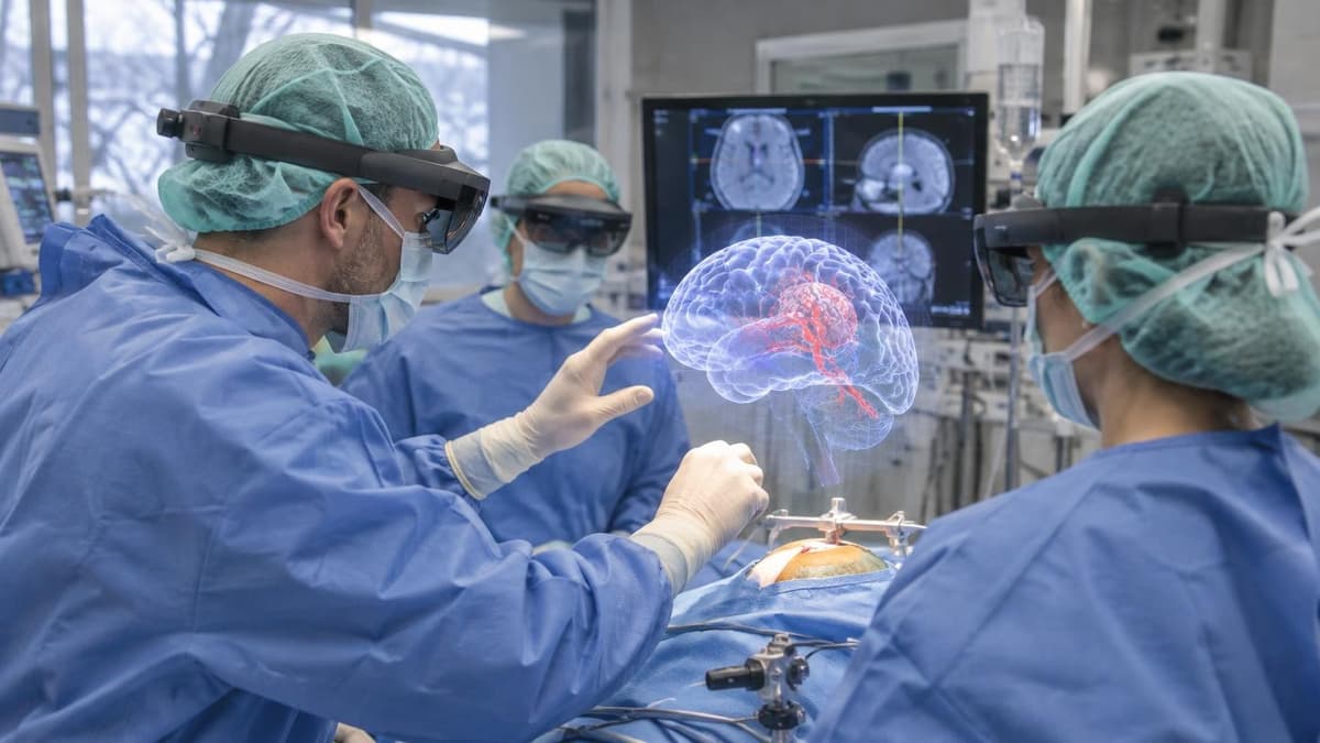
A three-dimensional brain image now appears above the operating table at Zurich University Hospital. For the first time, the hospital has used mixed-reality glasses to guide brain surgery, projecting a hologram based on the patient’s pre-operative computer tomography scan. The surgeon can view the brain’s structure while working, rather than relying only on landmarks visible from outside the head.
The procedure involved placing an external ventricular catheter, a thin tube that enters a brain ventricle to drain excess cerebrospinal fluid. The target lies deep inside the brain, making accurate placement difficult. The hospital said that even a difference of a few millimetres can affect the outcome.
The glasses bring the scan’s information into the surgeon’s view at the operating table. Zurich University Hospital says the system is intended to help identify where the catheter should go. The first operation marks the technology’s introduction into the hospital’s neurosurgical practice.
Guide the catheter to its target
The catheter’s target is hidden deep within the brain. Before the glasses were introduced, surgeons inserted the tube manually and used externally visible landmarks as reference points. The ventricular target itself cannot be seen from outside the patient’s head, Zurich University Hospital said.
The mixed-reality system displays data from a CT scan taken before the operation. Through the glasses, the surgeon sees a three-dimensional representation of the brain projected above the patient. The scan gives the surgical team a view of the internal structure while positioning the catheter, which must reach a ventricle to drain excess cerebrospinal fluid.
That added visual reference matters because the hospital says a few millimetres can determine the success of the procedure. The hologram therefore supports a task where the intended destination is internal and precise. It changes what the surgeon can see during placement, while the catheter remains a manually inserted tube.
Keep surgical judgement central
The glasses put CT information directly into the surgeon’s field of view. Menno Germans, a senior consultant in the hospital’s neurosurgery department, performed the operation. He said: “The information from the CT scan is displayed exactly where we need it during the procedure.”
The system links imaging taken before surgery with the view of the patient during the operation. That gives the surgeon a three-dimensional reference for locating the catheter’s destination, rather than depending solely on external landmarks. The hospital presents the technology as a way to improve patient safety in a procedure where a small placement difference can matter.
The hospital also stresses that the visual overlay does not make the surgical decisions. Its doctors have been trained to use the glasses, and the technology serves as a tool during the operation. Medical knowledge and experience remain central to the surgeon’s judgement about how to proceed.
Extend the tool to further procedures
Zurich University Hospital plans to use the technology in further procedures. Doctors at the clinic have trained to work with the mixed-reality glasses, following the hospital’s first operation with a holographic brain display. The next uses will extend the system beyond its initial procedure, although the hospital describes it as assistance for surgeons rather than a substitute for their expertise.
The approach brings pre-operative imaging into the operating room as a three-dimensional projection above the patient’s head. For catheter placement, that view can help the surgeon identify the target inside the brain. The hospital’s account of the operation centres on this practical use: giving clinicians a visual reference during a task that has traditionally relied on landmarks seen from outside the head.
For patients treated at the Zurich clinic, the technology is now part of the hospital’s developing surgical toolkit. Its role remains tied to the surgeon’s decisions throughout the procedure.